Accréditations reconnues
- Accréditation COFRAC Essais n° 1-1793 et 1-2290
- ISO/IEC 17025
- Nadcap MTL
SEM (Scanning Electron Microscopy) coupled with EDX (Energy-Dispersive X-ray Spectroscopy) is a powerful technique used in the laboratory to study the morphology and chemical composition of samples. The main principles of SEM-EDX analysis are:
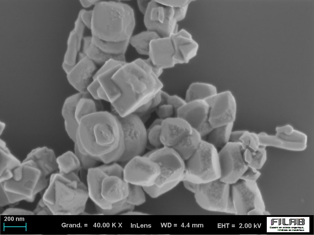
High-resolution imaging with SEM : SEM uses an electron beam scanned across the sample to produce an image of its surface. These images reveal the topography, morphology, and sometimes the chemical composition of the sample surface. SEM’s high resolution makes it possible to observe details at the nanometric scale.
Elemental analysis with EDX : EDX is often used in conjunction with FEG-SEM. When the SEM electron beam strikes the sample, it excites the atoms, causing them to emit characteristic X-rays. Analyzing these X-rays makes it possible to identify the chemical elements present in the sample and, in some cases, provide a quantitative dimension.
The FILAB analysis laboratory is now one of the leading French laboratories equipped with 3 SEMs.
This SEM microscopic analysis tool is particularly powerful and efficient for rapid diagnostics (contamination, inclusion, etc.) or more complex investigations.
At FILAB laboratory, SEM-EDX is the primary diagnostic tool. That is why we chose to integrate the following into our analytical equipment:
Two FEG-SEMs coupled with an EDX probe for materials failure analysis (metallic, polymer, ceramic, glass, etc.) and R&D.
A tungsten SEM coupled with an EDX probe for the identification of particles, contaminants, deposits and pollution.
SEM makes it possible to study different surfaces with great precision thanks to ultra high-definition image resolution
A variable pressure (VP) mode for insulating samples, enabling non-destructive analysis on all types of materials (without metallization)
An 80 mm² energy-dispersive microprobe (EDS), sensitive and fast, for performing semi-quantitative chemical analysis and mapping, including for light elements
An In-Lens detector for very high-resolution images of around 1 nm with accelerating voltages of about 1 kV
A transfer airlock allowing samples of «large size» to be introduced quickly and cleanly
At FILAB laboratory, we have a range of technical resources to carry out our chemical analysis successfully. The analytical coupling of SEM (Scanning Electron Microscope) with other analysis methods such as SEM-EDX, makes it possible to obtain detailed information on the chemical composition, crystalline structure and morphological properties of the samples observed. Thus, coupled SEM provides comprehensive characterization at the microscopic scale.
Our SEM-EDX expertise is fundamental to our analytical approach. At FILAB laboratory, our team of experts uses SEM-EDX analysis to meet complex analytical challenges, providing our clients with reliable, high-quality results across a range of services:
The identification of particles, contaminants and deposits
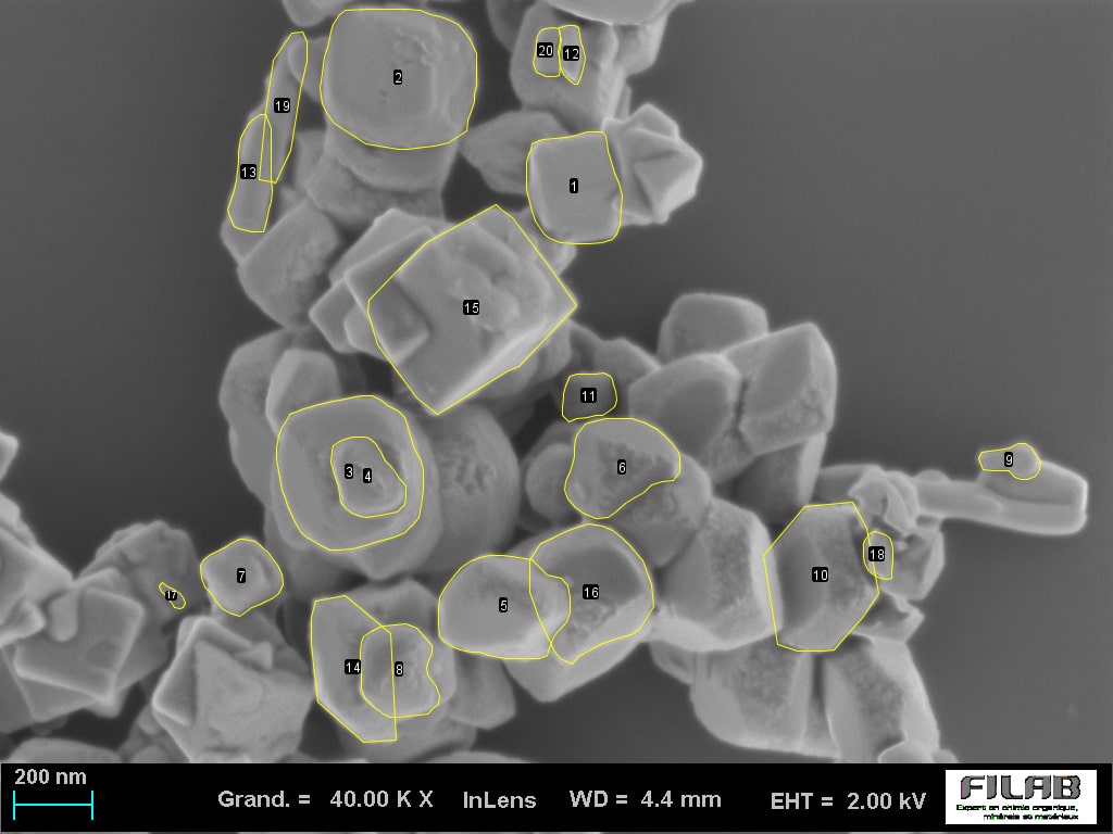
The nanometric characterization: FILAB is also the first French laboratory to be COFRAC ISO 17025 accredited in this field
Surface analysis
The combination of SEM images and EDX data provides a wealth of information about the sample, ranging from materials characterization to failure analysis. The results can reveal information about crystal structure, phase composition, element distribution, and other important properties of the samples studied, as part of a chemical analysis.
The Scanning Electron Microscopy technology SEM-EDX is a laboratory microscopic analysis technique using a field emission gun (FEG). This technique produces very high-resolution images of a sample’s surface (magnification of around *1000000). It is a technique used to study the chemical composition and morphology of solid materials. FILAB laboratory specializes in SEM-EDX expertise.
That is why FILAB laboratory decided to equip itself with not one, but 3 SEMs! Two are additionally coupled with EDX probes and one with an EBSD probe, enabling extreme precision in the analysis of your materials. These SEM-EDX techniques, equipped with in-column signal detection 20 times more intense than that of a conventional Scanning Electron Microscope, enable FILAB laboratory to carry out high-value-added analysis. Indeed, we obtain sharp images for non-destructive, precise, and much faster investigative observations and analysis.
Responsable expertise matériaux
Thomas Gautier accompagne les projets d’analyse MEB-EDX et de caractérisation des matériaux.
Responsable expertise matériaux
Thomas Rousseau accompagne les projets d’analyse MEB-EDX et de caractérisation des matériaux.
Transparency & contact Dernière vérification : juillet 2026
Ecoparc Dijon Bourgogne
80 rue Jean-Louis Auguste Petitjean
21850 Saint-Apollinaire, France
Tél. +33 (0)3 80 52 32 05
Request a MEB-EDX Quote"I used Filab for a chemical analysis. Very satisfied with the service. Responsive team, deadlines met, and a clear analysis report. I recommend them."
"Very responsive laboratory, with excellent analysis quality and deadlines met. Communication is smooth, the technical explanations are clear, and the team knows how to adapt to specific needs. I recommend them for their professionalism and reliability!"
"Clear and precise deliverable, delivered on time. I recommend them."
The SEM-EDX or SEM-FEG-EDX technique can be applied to all industrial fields, depending on the need for results:
An SEM-EDX analysis is a microscopy technique used for imaging and analyzing elements and chemical composition. This technique combines scanning electron microscopy (SEM) and energy-dispersive X-ray spectroscopy (EDX). SEM-EDX analysis makes it possible to observe surface structure and the chemical composition of materials at high magnification. The combination of these two techniques enables a detailed study of individual particles and the material’s surface features, which can be as small as 1 nanometer. The analytical information obtained from this analysis can provide valuable insights into the properties and characteristics of the sample in question. In addition, it can be used to identify contaminants or other impurities that may be present in trace amounts. SEM-EDX analysis is often used in a wide variety of applications, including materials science and forensic analysis. Thanks to this advanced technique, researchers can obtain precise information about the chemical composition of samples at a very small scale.
The process of SEM-EDX analysis begins with placing the sample in the chamber of an electron microscope. The electrons emitted by the sample then pass through an energy filter to produce secondary X-rays, which are detected by the EDX detector. The data relating to the energy-dispersed X-rays are then analyzed to determine the composition and concentration of the elements present in the sample. This analysis can provide precise results on particle size distribution, crystal structures, oxidation states, and other characteristics of individual particles.
SEM-EDX analysis is used across a wide range of industries, including materials science, aerospace engineering, medical research, electronics manufacturing, environmental studies, and forensic analysis. SEM analysis can be used to analyze and identify contaminants or other impurities present in trace amounts in materials and components. It can also help researchers better understand the surface characteristics of materials, such as particles or nanometer-scale structures. In addition, SEM-EDX analysis can provide valuable information on the composition and concentration of the elements present in samples for a variety of applications. Furthermore, this microscopy technique has been successfully used by the pharmaceutical industry to detect active ingredients in drug samples with great precision.
SEM-EDX analysis can provide several types of information:
The SEM-EDX analysis offers several advantages:
SEM-EDX analysis has a few limitations:
Interpreting the results of SEM-EDX analysis requires expertise and knowledge of the characteristics of the materials being studied. The results can be interpreted by comparing the energy spectra obtained with reference libraries containing characteristic spectra of known elements. The elemental maps and images obtained can be used to understand the spatial distribution of elements and to correlate microscopic observations with the chemical information obtained.