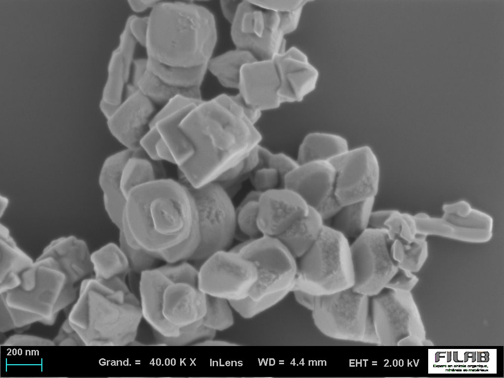
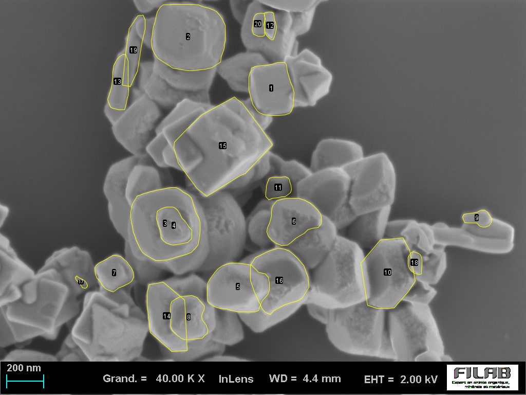
Scanning electron microscopy SEM-EDX in an analytical laboratory

What exactly is Scanning electron microscopy ?

Scanning Electron Microscopy (SEM-EDX) is an analytical technique which uses a Field Emission Gun (FEG) and produces high resolution images of a sample’s surface (1 000 000x zoom)

This new SEM-EDX with  column signal detection 20 times more powerful than a conventional Scanning Electron Microscope allows FILAB to carry out analyses with high added value and to get much clearer images for fast, non-destructive and accurate observations and investigations.

The FILAB analytical laboratory is today one of the first French laboratories to own a Zeiss GEMINI SEM microscope. This analytical tool is particularly powerful and efficient for rapidly diagnosing pollutants or for performing more complex expertise.

Discover examples of possible applications for a SEM-EDX microscope, specific to your field :

  • Fine chemistry / surface treatment
  • Luxury goods (Jewelry, Horology, Leathercraft…)
  • Cosmetics / pharmaceuticals / medical devices
  • Automobiles / aeronautics
  • Metallurgy / Nuclear energy

Take advantage of FILAB’s services and of the performance of a latest generation SEM-EDX-FEG :

  • An in-lens detector for high resolution pictures down to roughly 1nm with acceleration voltage of approximately 1kV.
  • A variable pressure mode (VP) for insulating samples to perform  non-destructive analysis on all types of materials (without metallization)
  • A transfer airlock which allows “large” samples to be rapidly and cleanly inserted
  • An 80mm² Energy-dispersive X-ray probe for performing semi-quantitative chemical analyses and  mapping even on lighter elements 

The positive aspects of FILAB

  • Technical expertise and extensive experience in many sectors of industry
  • Direct contact with one of our experts
  • Fast turnaround (24h priority service available)
  • Possibility to attend the analysis process
  • Competitive pricing

SEM-EDX analysis is a microscopy technique used for imaging and analysis of elements and chemical composition. This technique combines scanning electron microscopy (SEM) and energy dispersive X-ray spectroscopy (EDX). SEM-EDX analysis allows the surface structure and chemical composition of materials to be observed at high magnifications. The combination of these two techniques allows a detailed study of individual particles and surface characteristics of the material, which can be as small as 1 nanometre. The analytical information obtained from this analysis can provide valuable insights into the properties and characteristics of the sample in question. In addition, it can be used to identify contaminants or other impurities that may be present in trace amounts. SEM-EDX analysis is often used in a wide variety of applications, including materials science and forensic analysis. With this advanced technique, researchers can obtain precise information about the chemical composition of samples on a very small scale.


The SEM-EDX analysis process begins by placing the sample in the chamber of an electron microscope. The electrons emitted from the sample then pass through an energy filter to produce secondary X-rays which are detected by the EDX detector. The energy dispersed X-ray data is then analysed to determine the composition and concentration of elements present in the sample. This analysis can provide accurate results on particle size distribution, crystal structures, oxidation states and other characteristics of individual particles.

SEM-EDX analysis is used in a wide range of industries including materials science, aerospace engineering, medical research, electronics manufacturing, environmental studies and forensic analysis. SEM analysis can be used to analyse and identify contaminants or other trace impurities in materials and components. It can also help researchers to better understand the surface characteristics of materials, such as particles or nanoscale structures. In addition, SEM-EDX analysis can provide valuable information on the composition and concentration of elements present in samples for a variety of applications. In addition, this microscopy technique has been successfully used by the pharmaceutical industry to detect active ingredients in drug samples with high accuracy.

Thomas ROUSSEAU Scientific and Technical Director
Ask for your quote
+33 (0)3 80 52 32 05 Request
a quote
Thank you !

Click here to know more about our services

Logo Filab laboratoire analyse chimique